How AI is Transforming Radiology and Medical Imaging

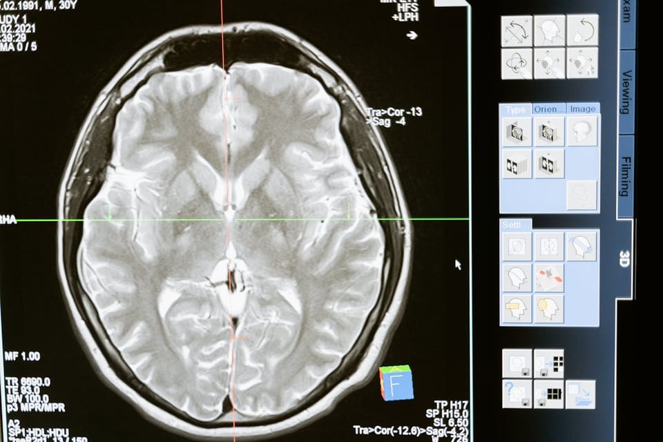
Table of Content
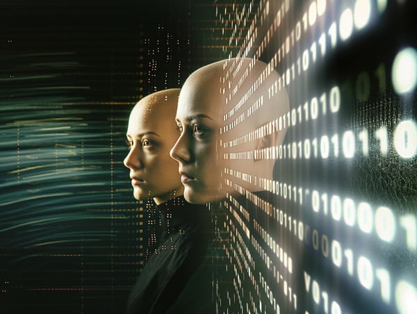
Artificial intelligence (AI) is revolutionizing industries around the world, and one of the fields most dramatically impacted by this technology is healthcare, particularly radiology and medical imaging.
AI's ability to process large amounts of data and identify patterns is enhancing the accuracy, speed, and efficiency of diagnostic imaging, enabling radiologists to better detect and treat diseases.
This transformation, especially with the rise of teleradiology solutions, allows artificial intelligence in radiology to make these advancements accessible remotely, significantly improving patient outcomes and reshaping how healthcare professionals approach radiological assessments, both in hospitals and through telemedicine platforms.
Keep reading to learn more about how AI is transforming radiology in today’s technology-driven world.
AI Enhances Accuracy in Diagnostics
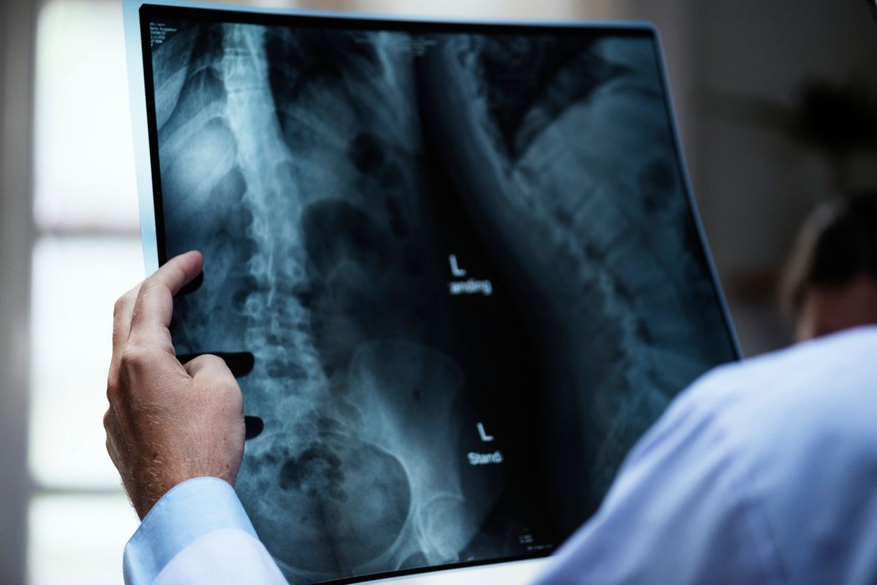
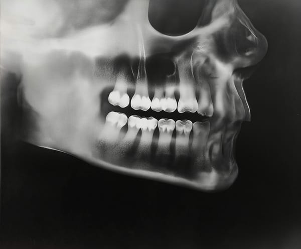
One of the key areas where AI is making a substantial difference in radiology is diagnostic accuracy. Medical imaging, such as X-rays, MRIs, and CT scans, generates complex data that radiologists traditionally analyze visually. Human interpretation, while skilled, can sometimes be subject to error or bias. AI, however, can assist by quickly analyzing these images and recognizing subtle patterns that may be missed by the human eye.
Deep learning algorithms, a subset of AI, are particularly effective at image recognition tasks. These systems are trained on large datasets of medical images and are capable of identifying specific features related to diseases such as cancer, fractures, and neurological disorders.
A great example of this is demonstrated in a 2020 study that showed AI models outperformed radiologists in detecting breast cancer from mammograms. The AI reduced false positives by 5.7% in the U.S. dataset and false negatives by 9.4%, improving diagnostic accuracy when used alongside radiologists.
Speed and Efficiency in Medical Imaging
Another area where AI is transforming radiology is in the speed and efficiency of image processing. Traditional methods of image analysis can be time-consuming, often requiring hours or days for detailed examination and interpretation. AI, however, can process vast amounts of data in a fraction of the time, providing quicker results without sacrificing accuracy.
This speed is especially valuable in emergency situations where time is critical. For instance, in the case of stroke patients, immediate and accurate identification of brain abnormalities is essential to begin life-saving treatments. AI algorithms can quickly analyze brain scans, identifying issues such as blood clots or hemorrhages in real time, allowing medical teams to respond faster and with greater confidence.
In addition, AI-driven automation in medical imaging helps reduce the workload for radiologists, who often face high volumes of cases daily. By automating routine tasks such as image sorting, measurements, and preliminary analysis, AI frees up radiologists to focus on more complex cases that require human expertise. This leads to increased efficiency in radiology departments and enables faster diagnosis for patients.
Personalizing Treatment Plans with AI
AI is not only enhancing how radiologists diagnose conditions but is also playing a pivotal role in personalizing treatment plans based on individual patient data. With the rise of teleradiology solutions, this capability extends even further, enabling radiologists and other healthcare providers to remotely access and analyze patient images from anywhere, facilitating real-time, personalized care regardless of location.
Medical imaging provides a wealth of information about a patient’s condition, and AI can integrate this data with other health records to offer a more comprehensive understanding of the disease, even across different medical facilities through telemedicine platforms.
Even more, AI’s ability to monitor changes in medical images over time enables ongoing, dynamic adjustments to treatment strategies. For example, in chronic diseases such as cardiovascular conditions, AI can analyze trends in imaging data to determine how a patient is responding to treatment, helping clinicians make timely modifications to improve outcomes.
AI in Early Disease Detection
Early detection of diseases is one of the most critical factors in improving patient survival rates, and AI is making significant strides in this area within radiology. By recognizing early-stage abnormalities in medical images, AI can detect diseases before they progress, allowing for earlier intervention and treatment.
One study shows that AI systems, namely machine learning and deep learning, have exhibited great success in predicting the potential occurrence of specific cancers and cardiovascular diseases. This research highlighted how AI excels at analyzing complex genetic data to accurately predict cardiovascular disease risk and identify unique genetic biomarkers, leading to more personalized treatment plans.
The Future of AI in Radiology
As AI technology continues to advance, its role in radiology and medical imaging will likely expand further. The integration of AI into everyday radiology practice will allow for faster, more accurate, and more personalized diagnoses, ultimately improving patient outcomes. Future developments in AI may include the ability to interpret increasingly complex types of medical data, further pushing the boundaries of what is possible in diagnostic medicine.
As AI continues to revolutionize radiology and medical imaging, doctors are becoming increasingly adept at using these technologies, applying their structured thinking and problem-solving skills to interact with AI systems—skills that make them natural prompt engineers in the evolving landscape of healthcare.
Overall, the collaboration between AI and radiologists has the potential to create a more efficient healthcare system, reducing costs and streamlining workflows. As AI becomes more integrated into medical imaging, patients can expect quicker, more accurate diagnoses, and treatments that are tailored to their specific needs.
 Hazem Abbas
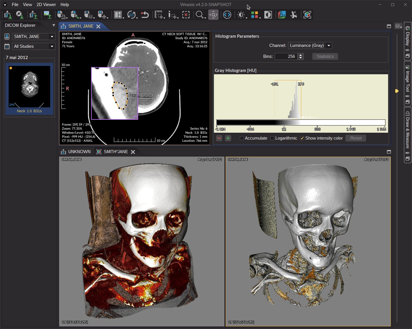 Desoky Mo
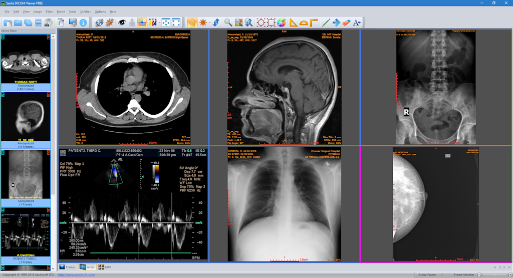 Hazem Abbas
 Desoky Mo
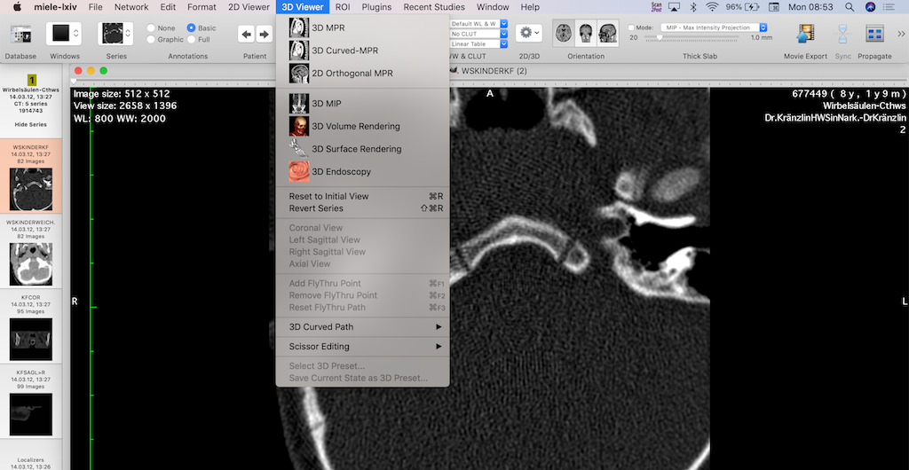 Hazem Abbas